If your orthodontist or dentist told you that your canine tooth never erupted properly, you’re definitely not alone. Impacted canines are more common than most people realize, and they’re often discovered by accident during routine X-rays or orthodontic evaluations.
When a canine tooth stays trapped in the bone instead of coming into place, it can start creating real problems over time. It may push against neighboring teeth, contribute to crowding, or interfere with proper bite alignment. In some cases, it can even lead to cyst formation around the impacted tooth. The surprising part is that many patients have no idea anything is wrong until imaging reveals it.
The key point is this: impacted canine treatment is not routine general dentistry. It requires surgical expertise. These cases are best managed by an oral and maxillofacial surgeon who regularly treats teeth that are unerupted, displaced, or positioned deep within the jawbone.
What Causes an Impacted Canine?
Canine teeth typically erupt around age 11 to 13. When they don’t, there is usually an underlying reason:
Lack of space: Crowding in the dental arch prevents normal eruption.
Retained baby tooth: A primary tooth may remain in place and block eruption.
Abnormal eruption path: The canine develops at an incorrect angle.
Genetics: Family history can increase the likelihood of impaction.
In many cases, the tooth is simply following a path that doesn’t bring it into the correct position. Patients often only learn about it after orthodontic evaluation or imaging.
How Impacted Canines Are Diagnosed
Impacted canines are not always visible during a clinical exam. They often sit high in the jaw or angled toward nearby tooth roots.
Diagnosis typically involves:
Standard dental X-rays to identify position
3D imaging (CBCT scans) for precise evaluation
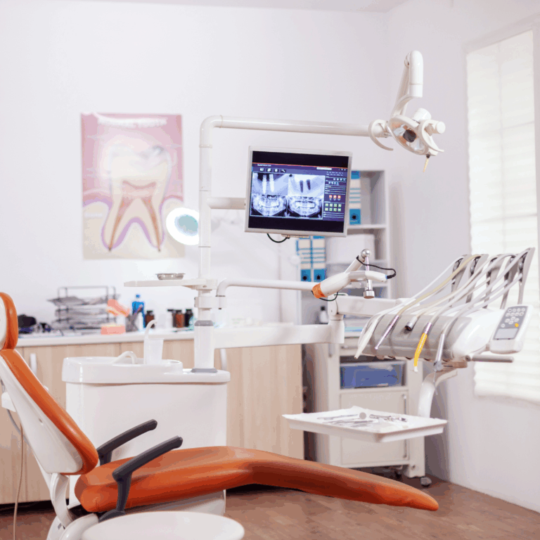
A 3D scan allows the oral surgeon to see the exact location, angle, and depth of the tooth, as well as its relationship to adjacent roots and nerves. This level of detail is critical for safe treatment planning and avoiding damage to healthy teeth.
At Somerset Oral Surgery, advanced imaging is used routinely to evaluate impacted teeth and determine the safest, most effective treatment approach.
Treatment Options for an Impacted Canine
There are two primary treatment paths, depending on the position of the tooth and overall orthodontic goals.
1. Surgical Exposure and Orthodontic Guidance
If the canine is in a favorable position, the goal is often to bring it into alignment rather than remove it.
In this procedure:
The oral surgeon exposes the impacted tooth
An orthodontic attachment is placed on the tooth
Braces or orthodontic traction gradually guide it into position
This is a controlled, gradual process that can successfully move the tooth into the correct place over time.
2. Surgical Extraction of the Impacted Tooth
If the canine is severely mispositioned or poses a risk to nearby teeth, removal may be the best option.
In these cases:
The tooth is surgically accessed through the gum and bone
It is carefully removed to protect surrounding structures
The area is allowed to heal before further orthodontic or restorative treatment
This approach is similar in complexity to other surgical extractions, including wisdom teeth removal, and is routinely performed by oral surgeons.
What the Procedure Is Like
Impacted canine surgery is typically performed under local anesthesia, with sedation options available for patient comfort.
During the procedure:
The area is fully numbed
Sedation helps patients remain relaxed or asleep
The surgeon completes the procedure with minimal discomfort
Most patients report feeling pressure rather than pain, and the procedure is generally well tolerated.
Recovery and Healing
Recovery depends on the complexity of the case and whether the tooth is being exposed or removed.
Typical healing timeline includes:
First 48–72 hours: Mild swelling and soreness are expected
3–5 days: Discomfort significantly improves
1–2 weeks: Soft tissue healing is well underway
Patients are usually advised to:
Eat soft foods initially
Use ice packs during the first 24 hours
Avoid strenuous activity for a short period
Follow all post-operative instructions carefully
If orthodontic traction is part of the plan, follow-up visits will continue to guide the tooth into position.
When to See an Oral Surgeon
You should consider evaluation if:
A canine tooth has not erupted by early teenage years
Your orthodontist suspects impaction
Imaging shows an unerupted or displaced canine
There is pressure, crowding, or bite changes in the area
Early evaluation allows for more predictable treatment options and can reduce the risk of damage to nearby teeth.
Final Thoughts
Impacted canines do not resolve on their own. Without treatment, they can continue to affect alignment, bite function, and surrounding teeth. The good news is that with proper diagnosis and surgical planning, outcomes are highly predictable.
An oral and maxillofacial surgeon can determine whether the tooth should be guided into place or removed safely, and coordinate closely with your orthodontist to ensure long-term stability.
If you are unsure about an impacted canine, a consultation at Somerset Oral Surgery can help clarify the situation and outline the best next steps for your case.
FAQ
How do I know if I have an impacted canine?
Most impacted canines are identified on dental X-rays or 3D scans rather than by symptoms alone.
Can an impacted canine come in naturally over time?
Once a canine is fully impacted, it is unlikely to erupt without surgical and orthodontic assistance.
Is surgical exposure painful?
The procedure is performed under anesthesia or sedation, and post-operative discomfort is typically mild and temporary.
Will I lose the tooth if it is impacted?
Not necessarily. Many impacted canines can be guided into position if they are in a favorable location.